
What to Do If You Suspect a Broken Bone: Fracture First Aid, X-Ray vs. CT & When to Go to the ER
Clinically reviewed by Mario Quintanilla, MD, Board-Certified Emergency Physician, on October 17, 2026.
Bellaire ER is a freestanding emergency room at 5302 Bellaire Blvd, open 24/7/365. Walk in or call ((713) 660-0001) anytime.
- Open now, 24/7/365
- Walk in, no appointment needed
- 5302 Bellaire Blvd, free parking
Clinical Triage Answer: Seek immediate emergency room evaluation if a suspected broken bone causes visible deformity or angulation, bone protruding through or tenting the skin, numbness or pale cold fingers or toes, or an inability to bear weight for four consecutive steps. Never attempt to straighten a deformed limb yourself. While plain X-rays detect most fractures, 15% to 20% of acute scaphoid wrist fractures and occult hip fractures do not appear on initial plain radiographs and require on-site multi-slice CT imaging or protective orthopedic splinting.
At Bellaire ER, board-certified emergency physicians evaluate acute orthopedic injuries 24/7 inside private exam suites equipped with digital X-ray, multi-slice helical CT, intravenous analgesia, and custom fiberglass and aluminum-foam splinting.
Sprain vs. Fracture: Why Movement Alone Cannot Rule Out a Break
One of the most common myths in emergency orthopedics is that if you can move a joint or wiggle your fingers and toes, the bone cannot be broken. In clinical reality, tendons and ligaments frequently remain continuous across hairline, non-displaced, impacted or pediatric buckle fractures. Patients regularly walk into the emergency department on fractured ankles or move their wrists despite an acute distal radius or scaphoid break.
In a landmark multicenter study published in JAMA by Dr. Ian Stiell and colleagues (PMID: 8114236) and confirmed in a systematic review of 15,581 patients in the BMJ by Dr. Lucas Bachmann and colleagues (PMID: 12595378), researchers established the Ottawa Ankle and Foot Rules. Their findings demonstrated that radiography is clinically required whenever an injured patient has bone tenderness along the posterior edge or tip of either ankle malleolus, tenderness at the base of the fifth metatarsal or navicular bone, or an inability to bear weight for four steps both immediately after the injury and in the emergency department. These criteria achieve a 98% to 99.6% sensitivity for detecting clinically significant fractures.
| Clinical Feature | Severe Ligament Sprain | Acute Bone Fracture (ICD-10 S52.509A / S82.899A) |
|---|---|---|
| Sound at Moment of Injury | Soft pop or tearing sensation | Audible snap, crack or grinding sensation (crepitus) |
| Location of Maximal Pain | Diffuse tenderness over soft tissue and ligaments | Pinpoint focal tenderness directly over the bone cortex |
| Weight-Bearing Ability | Often able to take 4 limping steps despite discomfort | Frequently unable to bear weight for 4 consecutive steps |
| Limb Contour | Generalized soft-tissue swelling around the joint | Visible angulation, shortening, rotation or step-off |
| Neurovascular Status | Normal distal pulse and sensation | Risk of distal numbness, tingling or diminished pulse |
| Definitive Diagnosis | Exclusion of fracture via multi-view X-ray or CT | Confirmed cortical disruption on X-ray or multi-slice CT |
Red Flag Symptoms: Call 911 or Go to the ER Immediately
Certain fracture presentations threaten limb viability, blood supply, or life and require immediate emergency department care rather than an urgent care clinic:
- Open (Compound) Fractures: Any cut, puncture or skin tear over the fracture site means the broken bone has communicated with the outside environment. In a landmark orthopedic trauma investigation published in the Journal of Orthopaedic Trauma by Dr. William Lack and colleagues (PMID: 25526095), administering intravenous antibiotics within 60 minutes of an open fracture reduced deep bone infection rates from 17.5% down to 6.8%.
- Visible Deformity or Joint Dislocation: A limb bent at an unnatural angle, shortened or rotated can compress or kink adjacent arteries and nerves until a physician performs a controlled reduction.
- Neurovascular Compromise (The “5 Ps”): Pain out of proportion to the injury, Pallor (pale or dusky fingers or toes), Paresthesia (pins-and-needles numbness), Paralysis, or Pulselessness indicate arterial compression or acute compartment syndrome, a surgical emergency where pressure inside the muscle fascia cuts off capillary blood flow.
- High-Energy Trauma or Axial Skeleton Injury: Suspected fractures of the hip, femur, pelvis, skull, neck or spine, or any injury following a motor vehicle collision or fall from height, require trauma bay stabilization and CT imaging.
- Elderly Fall with Hip or Groin Pain: Even if an older adult can still move their leg in bed after a slip, an impacted femoral neck or intertrochanteric hip fracture carries high morbidity if weight is placed on the joint.
Immediate First Aid: What to Do Before You Reach the ER
Proper pre-hospital care protects blood vessels, nerves and soft tissue while you travel to the emergency department:
- Immobilize the Limb in the Position Found: Support the injured arm or leg above and below the suspected break using a padded board, rolled magazine, pillow or sling. Never attempt to straighten, pull or push a deformed bone back into alignment. Forcing a displaced fracture without radiographic guidance and muscle relaxation can lacerate the radial, ulnar, tibial or popliteal artery.
- Control Bleeding and Cover Open Wounds: If skin is broken near the fracture, cover the wound gently with a sterile gauze pad or clean cloth. Apply gentle pressure around the edges to control bleeding, but never push directly on a protruding bone fragment.
- Apply Cold Therapy Indirectly: Wrap an ice pack or bag of ice in a towel and place it gently alongside the swollen area for 15 to 20 minutes at a time. Never apply ice directly to bare skin, which causes frostbite and masks nerve numbness.
- Remove Constriction Immediately: Slide off rings, watches, bracelets and tight footwear from the injured extremity right away. Once fracture edema peaks over the first two hours, a metal ring on a swollen finger can act as a tourniquet and cut off digital circulation.
- Nothing by Mouth (NPO): Do not eat solid food or drink liquids if the injury is severe or deformed. Keeping your stomach empty allows the emergency physician to safely administer intravenous procedural sedation for closed fracture reduction or coordinate immediate orthopedic surgery.
When Plain X-Rays Are Not Enough: Occult Fractures and Multi-Slice CT
Standard two-dimensional X-rays are the frontline diagnostic tool for most extremity fractures, but overlapping bone shadows and non-displaced trabecular cracks can hide serious breaks during the first 72 hours after trauma.
According to a comprehensive diagnostic review published in the Cochrane Database of Systematic Reviews by Dr. Wouter Mallee and colleagues (PMID: 26045406), approximately 15% to 20% of acute scaphoid wrist fractures after a fall onto an outstretched hand (FOOSH, ICD-10 S62.009A) appear completely normal on initial four-view wrist X-rays. Because the scaphoid bone receives its blood supply in a retrograde pattern through a tiny distal vessel, an untreated occult scaphoid fracture carries a high risk of avascular necrosis (bone death) and permanent wrist arthritis. Multi-slice computed tomography (CT) provides 99% specificity and superior cross-sectional detail to visualize hidden cortical fractures of the wrist, tibial plateau, ankle talus, calcaneus and hip on the same visit.
Pediatric Fractures: Growth Plates and Buckle Injuries
Children’s bones are more porous and flexible than adult bones, and they contain open epiphyseal growth plates (physes) near the ends of long bones. Instead of snapping cleanly across both sides of the bone shaft, pediatric falls often produce:
- Buckle (Torus) Fractures: One side of the bone cortex compresses and bulges outward without a complete break. In a randomized controlled trial of 113 children published in Pediatrics by Dr. Amy Plint and colleagues (PMID: 16510648), researchers demonstrated that low-risk pediatric distal radius and ulna buckle fractures treated with a removable orthopedic splint achieved faster return to normal physical function and higher patient comfort than rigid circumferential plaster casts.
- Greenstick Fractures: The bone bends and cracks through one cortex while the opposite side bows intact.
- Salter-Harris Growth Plate Fractures: Fractures passing through or across the growth plate require precise radiographic classification and orthopedic follow-up to prevent premature growth arrest or limb-length discrepancy.
How Bellaire ER Evaluates and Treats Suspected Fractures 24/7
Walking into a retail urgent care clinic with a severe fracture often results in a second bill and an ambulance transfer when the clinic discovers the break is displaced, open or requires CT imaging. At Bellaire ER, located at 5302 Bellaire Blvd, Bellaire, TX 77401, our freestanding emergency department provides hospital-level orthopedic trauma capabilities with Direct Private Exam Suite Intake:
- Immediate Pain Control and Neurovascular Assessment: Before positioning your limb for imaging, our nursing and physician team checks distal pulses, capillary refill, and two-point nerve sensation while providing targeted oral, intranasal, or intravenous analgesia so you are not left suffering during the workup.
- High-Resolution Digital X-Ray & Multi-Slice CT: Our on-site diagnostic imaging suite performs multi-angle digital radiography and high-resolution cross-sectional CT scans to identify intra-articular step-offs, occult scaphoid or hip fractures, and complex comminuted breaks.
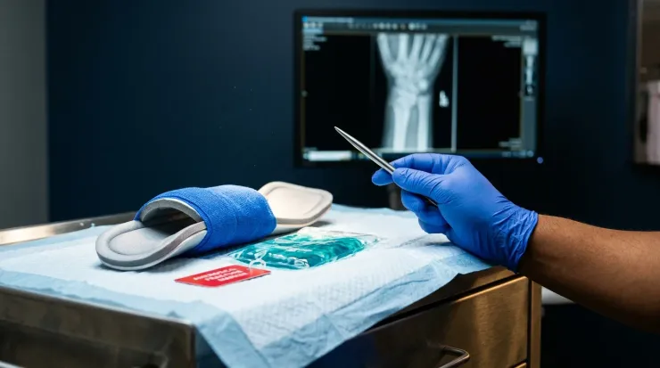
- Closed Reduction & Custom Splinting: During the first 48 to 72 hours after a fracture, soft-tissue swelling continues to expand. Placing a tight circular cast immediately can trap swelling and trigger compartment syndrome. Our emergency physicians apply custom-molded fiberglass or padded aluminum-foam orthopedic splints (
sugar-tong,ulnar gutter,thumb spica,posterior ankle), which immobilize the bone rigidly while accommodating acute swelling safely. - Open Fracture Stabilization & Direct Orthopedic Handoff: For open fractures, we administer immediate intravenous broad-spectrum antibiotics, tetanus prophylaxis, sterile high-volume wound irrigation, and coordinate direct orthopedic surgical consultation or inpatient hospital transfer when operative fixation (
ORIF) is required. - Coding & Clinical Documentation Reference: Common emergency diagnostic codes include
S52.509A(Unspecified fracture of the lower end of radius, initial encounter),S82.899A(Other fracture of lower leg/ankle, initial encounter),S62.009A(Unspecified fracture of navicular/scaphoid bone of wrist), andM84.40XA(Pathological fracture).
If you or your child suffered a fall, sports injury, or sudden impact in Bellaire, Meyerland, West University Place, or Galleria/Uptown, walk into Bellaire ER 24/7 or call (713) 660-0001 for immediate evaluation.
Frequently asked questions
Can you still move your fingers, toes, or joint if a bone is broken?
Yes. Being able to wiggle your fingers or toes or bend a joint does not rule out a fracture. Tendons and muscles often remain intact across non-displaced, hairline or buckle fractures, allowing partial movement despite a broken cortex. Only diagnostic X-ray or CT imaging can confirm or exclude a fracture.
How do doctors tell the difference between a bad sprain and a broken bone?
Emergency physicians use validated clinical decision criteria such as the Ottawa Ankle and Foot Rules alongside multi-view digital X-rays. Point tenderness directly over specific bone landmarks or the inability to bear weight for four consecutive steps carries a 98% to 99.6% sensitivity for identifying a fracture requiring radiography.
Can a broken bone be missed on a regular X-ray?
Yes. Approximately 15% to 20% of acute wrist scaphoid fractures, non-displaced femoral neck (hip) fractures in older adults, and subtle tibial plateau or calcaneal fractures are invisible on initial plain X-rays. When physical exam findings strongly suggest an occult fracture, on-site multi-slice CT scanning or precautionary splint immobilization is required.
Should I go to an emergency room or urgent care for a suspected broken bone?
Choose a 24/7 emergency room if the limb looks visibly deformed or crooked, bone is exposed through the skin, fingers or toes feel numb or pale, you cannot bear weight or walk, pain is severe, or you suspect a hip, pelvis, spine or complex joint fracture. Urgent care centers lack multi-slice CT scanners, intravenous pain control, and conscious sedation for closed fracture reduction.
What should I do immediately after a suspected fracture before reaching the ER?
Keep the injured limb completely still in the position you found it and never try to straighten or push a deformed bone back into place. Apply an ice pack wrapped in a cloth for 15 to 20 minutes, elevate the limb if doing so does not increase pain, cover any open skin wound with a clean sterile dressing, and withhold food or liquids in case sedation or surgery is needed.
Clinical references & peer-reviewed sources
- Implementation of the Ottawa Ankle Rules
- Accuracy of Ottawa ankle rules to exclude fractures of the ankle and mid-foot: systematic review
- Computed tomography versus magnetic resonance imaging versus bone scintigraphy for clinically suspected scaphoid fractures in patients with negative plain radiographs
- Type III Open Tibia Fractures: Immediate Antibiotic Prophylaxis Minimizes Infection
- A randomized, controlled trial of removable splinting versus casting for wrist buckle fractures in children
Have Questions About Visiting Bellaire ER?
Call our team at (713) 660-0001 any time with questions about visiting our independent, physician-owned freestanding emergency room at 5302 Bellaire Blvd, open 24/7 with short waits.